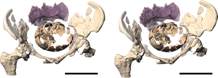
Vista anterior y superior de pelvis femenina de Homo sapiens en diferentes etapas del desarrollo (modificado de Huseynov et al., 2016).
El parto humano es un proceso que conocemos a la perfección cuando se trata de nuestra propia especie. Conocemos las diferentes fases del desarrollo y por supuesto somos conscientes de su dificultad. Una dificultad que radica precisamente en esas características que nos hacen diferentes, principalmente la bipedestación y la capacidad craneal. Nuestra historia evolutiva comenzó hace unos 7 millones de años, y consta de numerosos cambios que tuvieron como consecuencia la adaptación de las diferentes especies al entorno en el que vivían. Uno de los cambios más significativos fue la adquisición de la bipedestación. Un proceso que duró miles de años y que supuso numerosas variaciones anatómicas:
o La posición central del foramen magnum, respecto a la posición posterior en primates no bípedos.
o La adquisición de la forma de «S» en la columna vertebral, lo que permite repartir mejor el peso en el caso de la locomoción bípeda.
o La inclinación del fémur hacia la parte interna de las piernas.
o La alineación del dedo pulgar del pie respecto al resto de los dedos, lo que permite tener una mayor superficie de contacto al andar.
o Las variaciones en la pelvis, que hacen que sea una pelvis más ancha y corta que la de primates no bípedos.
Gracias a los descubrimientos realizados hasta la fecha, sabemos que estas adaptaciones anatómicas tuvieron consecuencias muy importantes para el parto humano. Las pelvis de especies pretéritas que se han conservado en el registro fósil nos permiten ahondar en algunos aspectos del parto de aquellos homininos. No se trata solamente del tamaño y de las características de la pelvis, ya que el tamaño del cráneo del feto también influye en el proceso del parto. Por desgracia, es muy complicado recuperar este tipo de restos, y más aún cuanto más atrás vamos en el tiempo. Por ello, muchas veces es necesario realizar complicados cálculos matemáticos para saber qué tamaño podrían tener los fetos y neonatos de los australopitecos y los primeros representantes del género Homo, entre otros.
¿Qué es lo que nos cuenta el registro fósil sobre el apasionante proceso del parto? Comenzaremos con uno de los esqueletos mejor conservados y conocidos por todos y todas: Lucy (individuo AL-288-1). Lucy era una hembra adulta de la especie Australopithecus afarensis que vivió al este de África hace unos 3,2 millones de años. Gracias a que se ha conservado en torno al 40% del esqueleto, podemos saber que se trataba de una especie bípeda, aunque mantenía ciertas características que le conferían una gran habilidad para moverse por los árboles. Probablemente, esta especie viviría en un entorno con densas masas boscosas, pero con grandes espacios abiertos entre ellas. Por lo tanto, se moverían por los árboles en busca de alimento, pero se verían «obligados» a caminar desde un espacio de bosque hasta el siguiente. La pelvis de Lucy nos indica que tenía un pubis alargado, muy parecido al de nuestra especie. Teniendo en cuenta que la capacidad craneal de esta especie estaba en torno a los 450 cm3, similar a la de los chimpancés, podríamos pensar que su canal de parto sería algo más holgado que en la actualidad. En lo que respecta al género Homo, se realizó el descubrimiento de una pelvis prácticamente completa en Gona (Etiopía) perteneciente a la especie Homo ergaster/Homo erectus que vivió hace 1,8 millones de años. Este hallazgo reavivó el debate sobre el parto en los homininos. Los investigadores encargados del estudio proponían que tanto los australopitecos como los primeros representantes del género Homo presentan una pelvis medio-lateralmente anchas, por lo que probablemente no tendrían un parto rotacional.
Vista frontal de la pelvis de un chimpancé (A), la pelvis del individuo AL-288-1 (Lucy, B), una pelvis femenina de Homo sapiens (C) y una pelvis masculina de Homo sapiens (D). Extraído de Lovejoy (2005).
Dentro del mundo de los neandertales, tenemos la suerte de que se hayan recuperado numerosos restos fósiles y generalmente en muy buen estado de conservación. No obstante, en algunos casos como el de la pelvis de Kebara 2, la deformación del sacro hizo proponer la hipótesis (que posteriormente se rechazó) de que los neandertales tenían un canal de parto estrecho y que por lo tanto hubieran tenido dificultades a la hora de dar a luz. Gracias a las técnicas más novedosas de tomografía computerizada hoy en día es posible realizar reconstrucciones en tres dimensiones con un alto nivel de fiabilidad. Los estudios realizados hasta la fecha sugieren que las hembras neandertales tendrían un canal de parto más ancho que las hembras de nuestra especie, por lo que podríamos pensar que tendrían una mayor facilidad a la hora de dar a luz. No obstante, la especie neandertal contaba con una media de capacidad craneal mayor que la del Homo sapiens. Se trataría, por lo tanto, de conocer el desarrollo del cerebro del feto para poder establecer la proporción entre el tamaño del cráneo y el tamaño del canal de parto en el momento del nacimiento. Para ello, se hicieron reconstrucciones en tres dimensiones del cráneo de un recién nacido neandertal (Mezmaiskaya, Rusia), de dos neonatos (Dederiyeh, Siria) y de la pelvis femenina de Tabun 1 (Israel). Los investigadores concluyeron que estos homininos ya tenían un parto rotacional (como en nuestra especie) y que se trataría de un parto de gran dificultad debido a la estrechez del canal pélvico con respecto al cráneo del bebé. Sin embargo, la hipótesis contraria fue propuesta por otro equipo de investigadores, sugiriendo que el canal de parto era lo suficientemente ancho como para que no fuese necesaria una rotación durante el nacimiento.
Reconstrucción del parto neandertal en base a la pelvis femenina de Tabun 1 y el cráneo de Mezmaiskaya (escala: 10 cm). Extraído de Ponce de León et al. (2008).
En conclusión, podemos decir que el parto es un tema apasionante no exento de debate y sobre el que se sigue investigando. No solamente es interesante conocer este proceso en especies pretéritas, sino también poner el foco en especies de primates actuales, como los chimpancés. Todo con el objetivo de arrojar luz sobre una cuestión que aún entraña muchas incógnitas. Como hemos podido observar, la clave de estos estudios es conocer tanto el tamaño de la pelvis como el tamaño del cráneo del bebé en el momento del nacimiento, ya que la relación entre ambos influirá en la mayor o menor dificultad del parto. A pesar de que la falta de conservación de los restos óseos en el registro fósil es un gran obstáculo, la esperanza de nuevos descubrimientos científicos y técnicas analíticas es un estupendo aliciente para la continuación de las investigaciones.
Bibliografía:
Huseynov, A., Zollikofer, C. P., Coudyzer, W., Gascho, D., Kellenberger, C., Hinzpeter, R., de León, M. S. P., 2016. Developmental evidence for obstetric adaptation of the human female pelvis. Proceedings of the National Academy of Sciences, 113(19), 5227-5232.
Lovejoy, C. O., 2005. The natural history of human gait and posture: Part 1. Spine and pelvis. Gait & posture, 21(1), 95-112.
Ponce de León, M. S., Golovanova, L., Doronichev, V., Romanova, G., Akazawa, T., Kondo, O., Ishida, H., Zollikofer, C. P., 2008. Neanderthal brain size at birth provides insights into the evolution of human life history. Proceedings of the National Academy of Sciences, 105(37), 13764-13768.
Ruff, C., 2010. Body size and body shape in early hominins–implications of the Gona pelvis. Journal of Human Evolution, 58(2), 166-178.
Simpson, S. W., Quade, J., Levin, N. E., Butler, R., Dupont-Nivet, G., Everett, M., Semaw, S., 2008. A female Homo erectus pelvis from Gona, Ethiopia. Science, 322(5904), 1089-1092.
Weaver, T. D., Hublin, J. J., 2009. Neandertal birth canal shape and the evolution of human childbirth. Proceedings of the National Academy of Sciences, 106(20), 8151-8156.
Zollikofer, C. P., Ponce de Leon, M. S., 2010. Reconsidering Neanderthal birth: Tabun mom, Kebara dad, Mezmaiskaya baby, and the consequences. In: American Journal of Physical Anthropology (pp. 252), Volume 141, Issue S50 (April 2010).
This translation was generated automatically using qwen2.5:14b-instruct and has not been reviewed by a human editor. It may contain errors, omissions or awkward phrasing — for the authoritative version, please read the original in Spanish. Found a mistake? Let us know at info@arqueotimes.es.
Anterior and superior view of female pelvis of Homo sapiens at different stages of development (modified from Huseynov et al., 2016).
Human childbirth is a process that we know very well when it comes to our own species. We understand the different stages of development and, of course, are aware of its difficulty. A difficulty that lies precisely in those characteristics that make us unique, mainly bipedalism and cranial capacity. Our evolutionary history began about 7 million years ago, and consists of numerous changes that led to the adaptation of various species to their environment. One of the most significant changes was the acquisition of bipedalism. A process that lasted thousands of years and resulted in numerous anatomical variations:
o The central position of the foramen magnum, compared to its posterior position in non-bipedal primates.
o The acquisition of an «S» shape in the spine, which allows for better weight distribution during bipedal locomotion.
o The inward tilt of the femur.
o The alignment of the big toe with respect to the other toes, allowing for greater surface area contact while walking.
o Variations in the pelvis, making it wider and shorter than that of non-bipedal primates.
Thanks to discoveries made so far, we know that these anatomical adaptations had very important consequences for human childbirth. The pelvises of prehistoric species preserved in the fossil record allow us to delve into some aspects of childbirth among those hominins. It is not just about the size and characteristics of the pelvis, as the size of the fetal skull also influences the birthing process. Unfortunately, it is very difficult to recover such remains, especially the further back we go in time. Therefore, many times complex mathematical calculations are necessary to determine what sizes the fetuses and neonates of australopithecines and early representatives of the genus Homo might have had.
What does the fossil record tell us about this fascinating process of childbirth? We will start with one of the best-preserved skeletons known to all: Lucy (individual AL-288-1). Lucy was an adult female of the species Australopithecus afarensis who lived in eastern Africa around 3.2 million years ago. Thanks to the preservation of about 40% of her skeleton, we know that she was a bipedal species, although she maintained certain characteristics that conferred great ability for moving through trees. Probably, this species would live in dense forest areas but with large open spaces between them. Therefore, they would move through the trees looking for food, but would be "forced" to walk from one wooded area to another. Lucy's pelvis indicates that it had an elongated pubis, very similar to ours. Considering that the cranial capacity of this species was around 450 cm3, similar to that of chimpanzees, we could think that her birth canal would be somewhat wider than today. As for the genus Homo, a nearly complete pelvis was discovered in Gona (Ethiopia) belonging to the species Homo ergaster/Homo erectus that lived 1.8 million years ago. This discovery reignited debate about childbirth among hominins. The researchers responsible for the study proposed that both australopithecines and early representatives of the genus Homo have pelvises that are moderately wide, so they probably would not have rotational births.
Frontal view of a chimpanzee pelvis (A), the pelvis of individual AL-288-1 (Lucy, B), a female Homo sapiens pelvis (C) and a male Homo sapiens pelvis (D). Adapted from Lovejoy (2005).
Within the world of Neanderthals, we are fortunate that numerous fossil remains have been recovered and generally in very good condition. However, in some cases like the pelvis of Kebara 2, sacral deformation led to the hypothesis (which was later rejected) that Neanderthals had a narrow birth canal and would therefore have difficulty giving birth. Thanks to the most innovative CT techniques today, it is possible to perform three-dimensional reconstructions with a high level of reliability. Studies conducted so far suggest that female Neanderthals would have a wider birth canal than those of our species, which could mean they had greater ease in childbirth. However, the Neanderthal species had an average cranial capacity larger than Homo sapiens. It would therefore be necessary to know the development of the fetal brain to establish the proportion between skull size and birth canal size at the time of birth. For this purpose, three-dimensional reconstructions were made of a newborn Neanderthal's (Mezmaiskaya, Russia) cranium, two neonates' (Dederiyeh, Syria), and the female pelvis of Tabun 1 (Israel). The researchers concluded that these hominins already had rotational births (like ours) and would have been difficult due to the narrowness of the pelvic canal compared to the baby's skull. However, another team of researchers proposed an opposing hypothesis suggesting that the birth canal was wide enough for a rotation not to be necessary during delivery.
Reconstruction of Neanderthal childbirth based on the female pelvis of Tabun 1 and the cranium of Mezmaiskaya (scale: 10 cm). Adapted from Ponce de León et al. (2008).
In conclusion, we can say that childbirth is an intriguing topic not without debate and one that continues to be researched. It is not only interesting to know this process in prehistoric species but also to focus on current primate species such as chimpanzees. All with the aim of shedding light on a question that still entails many uncertainties. As we have observed, the key to these studies lies in knowing both the size of the pelvis and the size of the baby's skull at birth, since their relationship will influence the greater or lesser difficulty of childbirth. Despite the lack of preservation of bone remains in the fossil record being a great obstacle, the hope for new scientific discoveries and analytical techniques is an excellent incentive for continuing research.
Bibliography:
Huseynov, A., Zollikofer, C. P., Coudyzer, W., Gascho, D., Kellenberger, C., Hinzpeter, R., de León, M. S. P., 2016. Developmental evidence for obstetric adaptation of the human female pelvis. Proceedings of the National Academy of Sciences, 113(19), 5227-5232.
Lovejoy, C. O., 2005. The natural history of human gait and posture: Part 1. Spine and pelvis. Gait & Posture, 21(1), 95-112.
Ponce de León, M. S., Golovanova, L., Doronichev, V., Romanova, G., Akazawa, T., Kondo, O., Ishida, H., Zollikofer, C. P., 2008. Neanderthal brain size at birth provides insights into the evolution of human life history. Proceedings of the National Academy of Sciences, 105(37), 13764-13768.
Ruff, C., 2010. Body size and body shape in early hominins–implications of the Gona pelvis. Journal of Human Evolution, 58(2), 166-178.
Simpson, S. W., Quade, J., Levin, N. E., Butler, R., Dupont-Nivet, G., Everett, M., Semaw, S., 2008. A female Homo erectus pelvis from Gona, Ethiopia. Science, 322(5904), 1089-1092.
Weaver, T. D., Hublin, J. J., 2009. Neandertal birth canal shape and the evolution of human childbirth. Proceedings of the National Academy of Sciences, 106(20), 8151-8156.
Zollikofer, C. P., Ponce de Leon, M. S., 2010. Reconsidering Neanderthal birth: Tabun mom, Kebara dad, Mezmaiskaya baby, and the consequences. In: American Journal of Physical Anthropology (pp. 252), Volume 141, Issue S50 (April 2010).
Alonso Caño, G. (2022, 10 de febrero). La prehistoria en clave femenina: algunos aspectos aspectos biológicos sobre el parto.. ArqueoTimes. https://arqueotimes.com/articulos/la-prehistoria-en-clave-femenina-algunos-aspectos-aspectos-biologicos-sobre-el-parto/





Comentarios